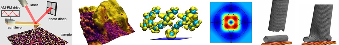
The major research thrusts in our program include (1) multifrequency AFM, (2) computational modeling of AFM, (3) fundamental developments in the characterization of viscoelatic behaviors involving multiple retardation times through AFM, and (4) nanoscale characterization of energy materials. Although we study a variety of topics, most of our projects are closely related to at least one of these thrusts.
1. Multifrequency AFM
With the introduction of the first bimodal AFM method by the Garcia group (Appl. Phys. Lett. 2004, Vol. 84, p. 449), a new family of techniques was born, which involves the simultaneous excitation of the AFM cantilever probe at more than one frequency, for the purpose of expanding the number of information channels that the user can acquire during a single 2-dimensional sample scan. The various excitation frequencies can be used to drive either a single cantilever eigenmode or multiple eigenmodes. Our objective is to develop new techniques that allow the user to simultaneously perform multiple characterization tasks, primarily through the excitation of multiple cantilever eigenmodes. A recent example of this type of development is trimodal AFM (ACS Nano 2013, Vol. 7, p. 10387), which allows simultaneous topographical imaging, compositional mapping and control of the tip-sample indentation during imaging, thus offering new capabilities for the subsurface visualization of soft matter. Our current focus is the development of tetra- and pentamodal AFM methods (Beilstein J. Nanotech. 2014, Vol. 5, p. 1637) for the study of viscoelastic materials that have multiple characteristic times (see also project # 3, below).
2. Computational Modeling of AFM
We dedicate a significant portion of our efforts to investigate, through computational modeling, the subtleties involved in the AFM measurement process itself, which are often neglected during routine characterization. This is useful in terms of correctly interpreting experimental results, in terms of predicting behaviors and capabilities for future experimental verification or implementation, and in terms of identifying challenges and complexities that offer new research opportunities. We have modeled a variety of AFM methods, including amplitude-modulation, frequency-modulation, various modes of multifrequency AFM (ACS Nano 2013, Vol. 7, p. 10387; Measurement Sci. & Technol. 2010, Vol. 21, No. 125502), band excitation (Nanotechnology 2012, Vol. 23, No. 015706; Small 2012, Vol. 8, p. 1264) spectral inversion (Nanotechnology 2010, Vol. 21, No. 075702), and single-impact viscoelastic characterization (based on band excitation, Sci. Rep. 2018, Vol. 8, p. 7534; Sci. Rep. 2019, Vol. 9, p. 12721), and often combine classical modeling with computational quantum mechanics (Nano Lett. 2011, Vol. 11, p. 5026) or molecular dynamics (J. Phys. Chem. B 2005, Vol. 109, p. 11493).
3. Fundamental Developments in the Characterization of Viscoelatic Behaviors Involving Multiple Retardation Times through AFM
Numerous emerging nanoscale technologies are based on viscoelastic materials, such as polymers and biological structures, whose behavior depends on their prior deformation history. Exploration of these materials with scanning probe microscopy methods can lead to a deeper understanding of the relationships between their nanomechanical properties and their performance in actual scientific and engineering applications. We are thus interested in developing routes towards the measurement of meaningful viscoelastic quantities (e.g., retardation times, frequency dependent loss angle and moduli, time dependent compliance, etc.) from AFM data, through existing experimental procedures such as tapping mode AFM, static force spectroscopy, and multifrequency AFM. Our approach involves (i) the development of tailored mathematical expressions and numerical fitting techniques based on rigorous models, such as the generalized Maxwell viscoelastic model (J. Pol. Sci B: Pol. Phys. 2017, Vol. 55, p. 804), (ii) computational modeling of realistic time-intensive viscoelastic behaviors (Beilstein J. Nanotech. 2016, Vol. 7, p. 554; Beilstein J. Nanotech. 2014, Vol. 5, p. 2149), and (iii) experimental validation on systems of interest such as polymers (Beilstein J. Nanotech. 2020, Vol. 11, p. 922) and biological systems (Nanoscale 2019, Vol. 11, p. 8918).
4. Nanoscale Characterization of Energy Materials
As a materials science initiative, we study ion exchange polymers and organic photovoltaic devices in order to better understand their functional and morphological changes as a function of environmental conditions, including chemical environment, humidity, temperature and mechanical stresses. We are especially interested in accelerated degradation studies that provide insight into failure mechanisms for fuel cells and solar cells. Our energy material studies are often closely related to the development of new AFM techniques, such as the proposed intermittent-contact conductive AFM (Beilstein J. Nanotech. 2020, Vol. 11, p. 453; also previously proposed by others in different form) and the development of procedures for characterizing electromechanical properties (Beilstein J. Nanotech. 2017, Vol. 8, p. 579; J. Appl. Phys. 2016, Vol. 119, No. 165301; J. Appl. Phys. 2014, Vol. 116, No. 104901; Beilstein J. Nanotech. 2014, Vol. 5, p. 1144).